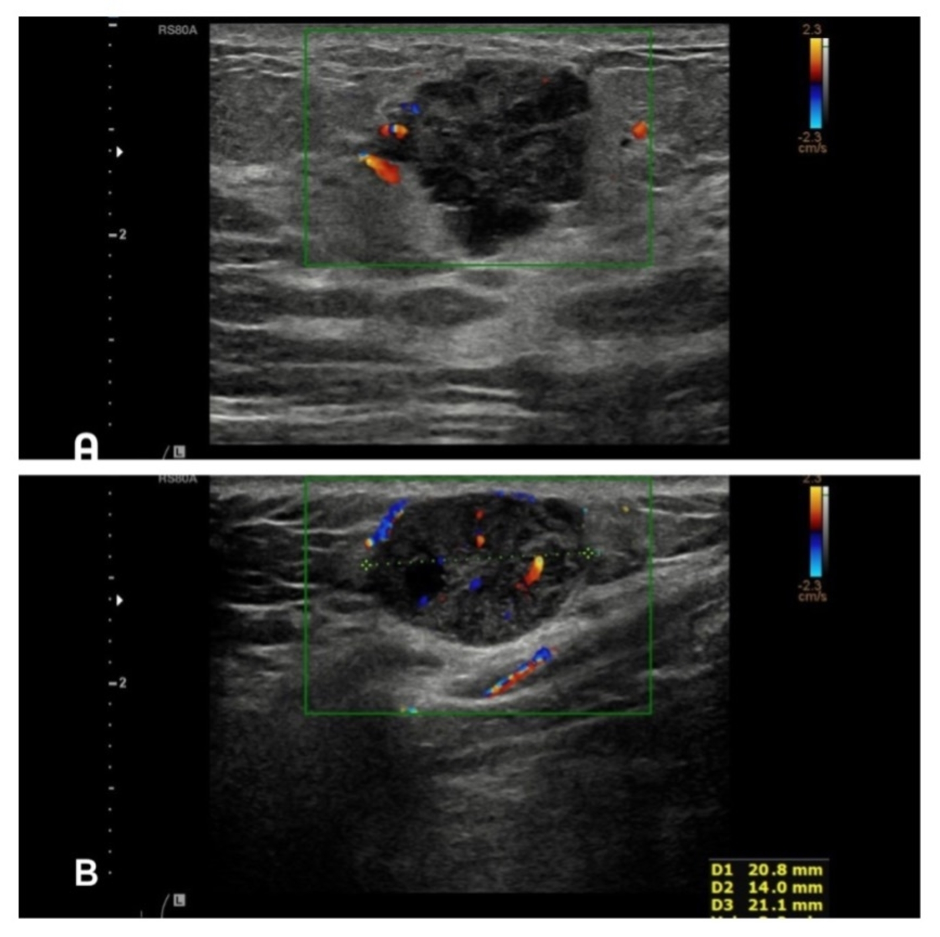
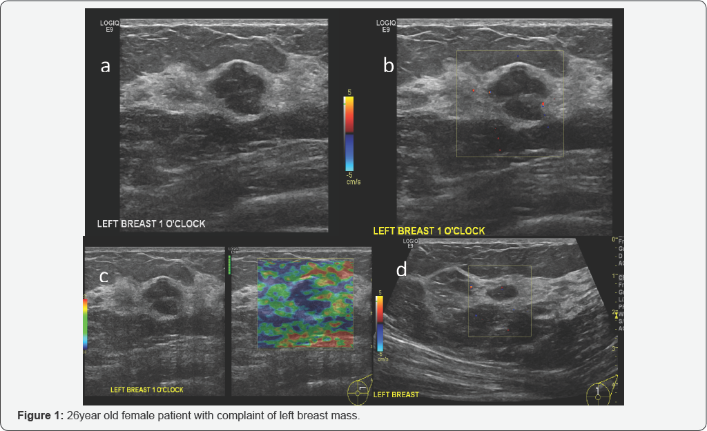
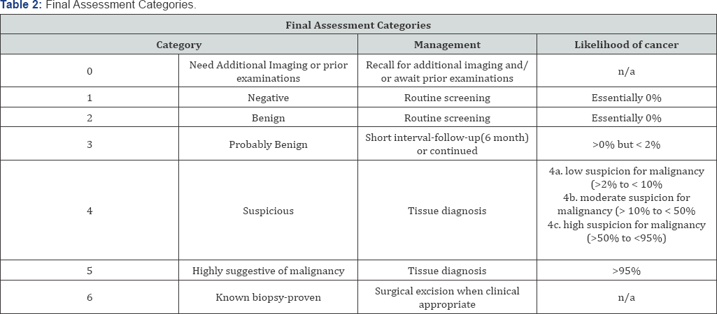
BREAST MASS DISCOVERED WITH CT SCAN UPDATE The breast cancer specialist and her PA both agree it's probably cancer, about 90% sure Will get scheduled for a mammogram and biopsy, ASAP Not smooth, like my gyn/onc told me, but irregular, vertical, 1/2", but movable No indications in lymph nodes, but will check those as well 0 #4 Breast quadrants According to SuperCodercom "The correct codes for the 12 o'clock position breast mass are N6322 and N6321 or N6311 and N6312 as per laterality mentioned in document" Another post from from terrilynnlogan@gmailcom states "There was a WPS call back on Jan 10th where this issue was addressedThree hypoechoic nodules at 3 o'clock position measures about 24 x 10 mm, 10 x 3 mm and 7 x 4 mm (d)with no internal vascularity in Doppler exam (not shown) (Figure 2) ImpressionCase 2 Abovedescribed finding representing bilateral breast fibro adenomas BIRADS 3
Cureus Metastatic Malignant Melanoma With Occult Primary Presenting As Breast Mass A Case Report And Literature Review
Left breast cancer 9 o'clock position icd 10
Left breast cancer 9 o'clock position icd 10- Tumor size is an important factor in breast cancer staging, and it can affect a person's treatment options and outlook Tumors are likely to be smaller when doctors detect them early, which can3 o'clock position (right breast) is towards the middle of your chest3 o'clock position (left breast ) is towards your armpitHope I've been helpfulRegards Comment japdip Breast cancer is not an inevitability From what you eat and drink to how much you exercise, learn what you can do to slash your risk


Icd 10 Code For Breast Cancer At 12 00
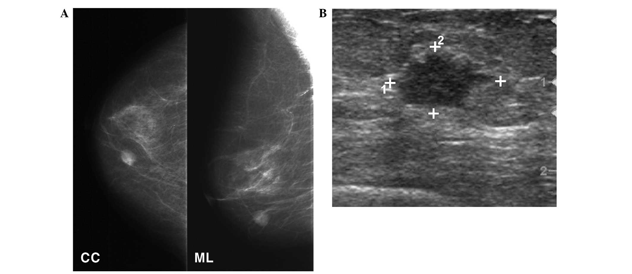
The second most common type of breast cancer is invasive lobular carcinoma, which accounts for 5%–10% of all malignant breast tumors (, 10) Bilateral craniocaudal mammograms reveal a focal asymmetric density at the 12 o'clock position in the right breast (arrow) Additional mammography and US were performed due to suspected occult When the breast is lifted up to position it on the image receptor, the tissue along the periphery of the breast should be palpated and additional special views of any thickening or mass that would not be imaged on the routine twoview mammogram should be obtained 30° Superolateralto 60° MediolateralThere is on benign appearing solid lesion at 10 o'clock position in the (R) breast measuring from the nipple This is stable since the previous study In the (L) breast, there is a solid lesion at 4 o'clock position, 5 cm from the nipple It has microlobulated margins and
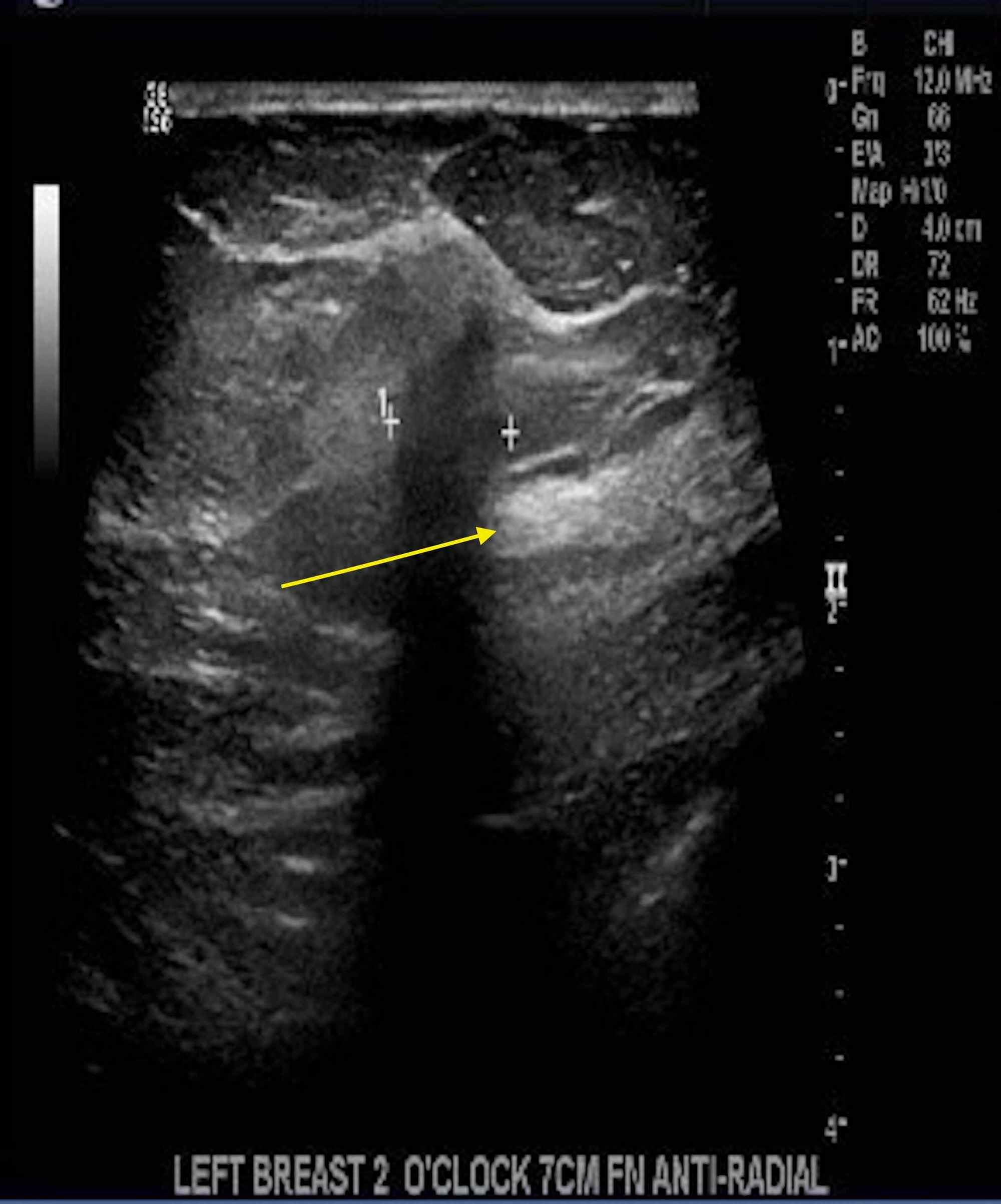
ICD 10 AM Edition Eleventh Edition Query Number 3598 In the Eleventh Edition Education slides Neoplasms, there is a diagram with clock positions with an example of documentation of a breast lesion at 6 o'clock coded to C508 Overlapping lesion of breast VICC #3107 Breast Quadrants and neoplasm codes advises to use a diagram as a reference to assistMammogram Circumscribed mass in the lower inner left breast which persists on spot compression views (not shown) Ultrasound Echogenic solid mass with marked vascular flow on color Doppler imaging at 8 o'clock 10 cm from the nipple measuring 6 x 4 x 6 mm, corresponding to the site of the mammographic massCase Discussion MACROSCOPIC DESCRIPTION "Left breast 1 o'clock, 7cm FN" Four cores 30mm in total length A1 MICROSCOPIC DESCRIPTION Sections show multiple cores of breast parenchyma which shows an invasive carcinoma with morphological features in keeping with a BRE grade 2 tumor DIAGNOSIS Left breast 1 o'clock 7cm FN Invasive carcinoma NST
With premenopausal breast cancer Digital mammography shows area of calcifications Magnification views demonstrate intraductal pleomorphic microcalcifications in the right 11 o'clock area Final diagnosis indicated intraductal carcinoma in situ D0511 Intraductal carcinoma in situ of right breast Malignant neoplasm of upperouter quadrant of right female breast 16 17 18 19 21 Billable/Specific Code C is a billable/specific ICD10CM code On physical exam, a 10mm mass was palpated in the left breast at the 10 o'clock position, medial to the areola A screening mammogram performed four months earlier demonstrated scattered fibroglandular densities bilaterally with mild stable architectural distortion in the right retroareolar region at the site of prior benign lumpectomy, and bilateral benign


Cureus Metastatic Malignant Melanoma With Occult Primary Presenting As Breast Mass A Case Report And Literature Review


Icd 10 Cm Coding Overlapping Breast Quadrants Coffee With A Medical Coding Auditor
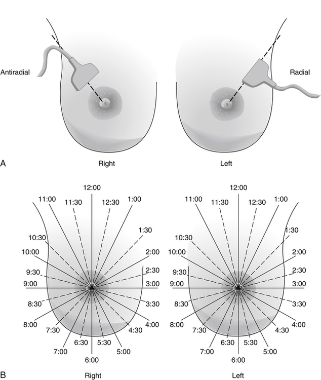
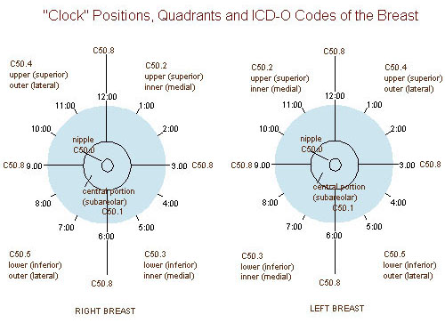
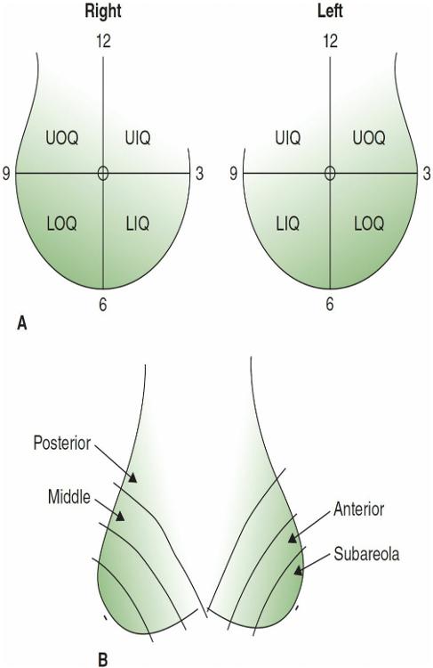
November last year a lump was discovered on my left breast at the 9 o'clock position Had it biopsied and it was determined to be fat necrosis Three weeks ago I found what seems to be a hard lump around the 9 o'clock position I had a follow up Manmo and ultrasound in may of this year and nothing showedThe following classification is recommended by the National Breast Cancer Centre and endorsed by the Royal Australian and New Zealand College of Radiologists1 Figure 6c Ultrasound confirms an irregular mass lesion with indistinct margins at the 12 o'clock position, 100 mm from the nipple Hyperechoic foci within the lesion Clock face description of breast lesion locations The clock face location of breast findings is described by imaging a clock on both the left and the right breast as the woman faces the examiner Note that the outer portion of the breast on the right is at the 9o'clock position and the outer portion on the left is at the 3o'clock position


Methylene Blue Dye Related Changes In The Breast After Sentinel Lymph Node Localization Kang 11 Journal Of Ultrasound In Medicine Wiley Online Library


Undiagnosed Breast Cancer Features At Supplemental Screening Us Radiology
Fig 61 At the 12 o'clock position of the left breast and the 3 o'clock position of the right breast, there are two oval masses (arrows) The spot compression views demonstrate that the margins of the right mass are well defined, and the margins of the left mass are ill defined (A) Right MLO mammogram (B) Left MLO mammogramIn T1b, the tumor size is greater than 5 mm, but less than or equal to 10 mm T1a and T1b breast cancers without lymph node spread have excellent longterm outcomes, with more than 95% of women alive at 10 yearsFamily history should note breast cancer in a 1stdegree relative (mother, sister, daughter) and, if family history is positive, whether the relative carried one of the inherited gene mutations that predispose to breast cancer (eg, BRCA1, BRC) Physical examination


2


Usefulness Of 3 Dimensional Printed Breast Surgical Guides For Undetectable Ductal Carcinoma In Situ On Ultrasonography A Report Of 2 Cases
The margin is irregular Another three hypoechoic nodules noted at 8 and 10 o'clock position of right breast and behind the nipple (05 x 07 cm, 05 x 05 cm and 17 x 18 cm) A right axillary node is also seen, 09 x 08 cm Impression Findings in keeping with Ca breast In T1a breast cancer, the tumor size is less than or equal to 5 millimeters (mm); The imaging of the right breast is normal ( not shown ) The left breast demonstrates a 1cm isodense oval mass in the posterior depth of the left breast at the 6 to 7 o'clock location, 10 cm from the nipple ( Fig 708, Fig 709, Fig 7010 ) A fatty hilum is suggested on the lateromedial (LM) spotcompression digital breast tomosynthesis


Breast Cancer Topic If You Are Waiting Please Let Me To Share My Experience


Q Tbn And9gcth39qrs02wrthtpk Uisqk0p0rnefiz Yi0mmwn2cv4eblwxg Usqp Cau
Posterior breast cancer mammographic and ultrasonographic features Vojnosanit Pregl 13 Nov;70(11) doi /vspj Authors Ana Janković 1Code C509 (Breast, NOS) in this situation Code the primary site to C508 when • there is a single tumor in two or more subsites and the subsite in which the tumor originated is unknown • there is a single tumor located at the 12, 3, 6, or 9 o'clock position on the breastThere is a 7mm hypoechoic nodule with shadowing at the 10 o'clock position of the right breast The biopsy sampling of the ill defined opacity in the upper outer right breast on mammography Also, biopsy of the 7mm hypoechoic solid nodule at the 10 o'clock position in the right breast, only seen with sonography Birads category 4


Figure 2 From Efficacy Of Pectoral Nerve Block Type Ii For Breast Conserving Surgery And Sentinel Lymph Node Biopsy A Prospective Randomized Controlled Study Semantic Scholar


Breast Mass Radiology Key
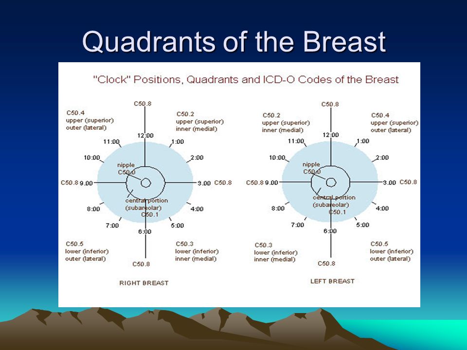
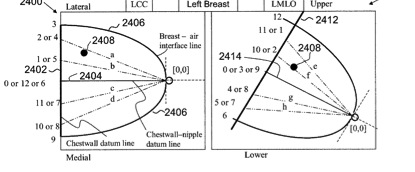
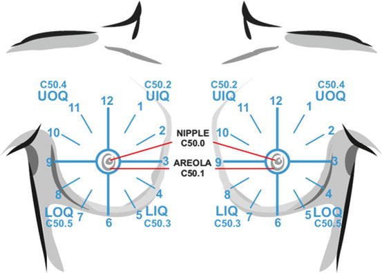
Code C509 (Breast, NOS) in this situation Code the primary site to C508 when O'Clock Positions and Codes Quadrants of Breasts 2 11 12 1 1 10 9 8 7 7 6 5 4 3 2 11 12 10 6 5 3 RIGHT BREAST LEF T BREAST UOQ UIQ UIQ UOQ LOQ LIQ LIQ LOQ C504 Given this guidance, a 12 o'clock right breast mass can be reported as ICD10 code N6315, Unspecified lump in right breast, overlapping quadrants, or as dual ICD10 codes for overlapping quadrants, N6311, Unspecified lump in the right breast, upper outer quadrant, and N6312, Unspecified lump in the right breast, upper inner quadrantCode C509 (Breast, NOS) in this situation O'Clock Positions and Codes Quadrants of Breasts 2 11 12 1 1 10 9 8 7 6 5 7 4 3 2 11 12 10 6 5 4 3 RIGHT BREAST LEF T BREAST UOQ UIQ UIQ UOQ LOQ LIQ LIQ LOQ C504 C502 C502 C504 C505 C503 C505 C503 C500 C501


Spie Org Samples Pm211 Pdf


The Breast Cancer Warriors Of Pakistan Posts Facebook
One lump was aspirated (upper inner right breast marked by arrows in A and B), yielding malignant cytology (D) Repeat ultrasonography of the known malignant mass in the right breast at 2o'clock position shows it to be indistinctly marginated and lobulated both without and (E) with spatial compounding Any lesion documented on Breast cancer, ultrasonography Mediolateral oblique digital mammogram of the right breast in a 66yearold woman with a new, opaque, irregular mass approximately 1 cm in diameter The mass has spiculated margins in the middle third of the right breast at the 10o'clock positionThe outer left breast is at 3 o'clock and the outer right breast is at 9 o'clock In the left breast the upper outer quadrant is between 12 and 3 o'clock The radiologist will also describe the size and location of a finding by indicating the distance from the nipple in centimeters


Missed Breast Cancer Effects Of Subconscious Bias And Lesion Characteristics Radiographics


Breast Cancer Signs Symptoms And Understanding An Imaging Report Saint John S Cancer Institute
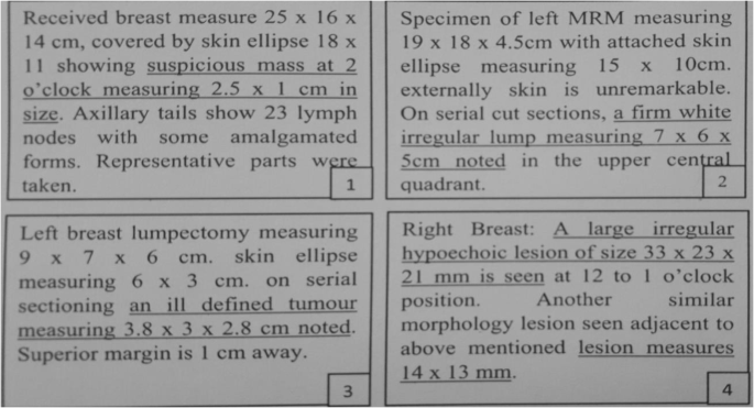
Pathology The most common cause for an asymmetry on screening mammography is superimposition of normal breast tissue (summation artifact) 6Asymmetries that are subsequently confirmed to be a real lesion may represent a focal asymmetry or mass, for which it is important to further evaluate to exclude breast cancer 5Developing asymmetries are sufficiently suspiciousABUS detected a right breast irregular hypoechoic mass with angular margins and posterior acoustic shadowing at the 1030 o'clock position, measuring 15 x 16 x 11 cm, 7 cm from the nipple Dedicated handheld ultrasound confirmed the mass A core biopsy was performed and detected invasive moderately differentiated ductal carcinoma On 14 March 14, WF had an ultrasound of her breasts "There is a 17 mm x 96 mm lesion at 2 o'clock position of left breast, 4 cm from the nipple" A FNAC (Fine needle aspiration cytology) done in a Taiping private hospital showed "benign breast


Missed Breast Cancer Effects Of Subconscious Bias And Lesion Characteristics Radiographics


Anatomic Stage Extraction From Medical Reports Of Breast Cancer Patients Using Natural Language Processing Springerlink
Lumpectomy (lumPEKtuhme) is surgery to remove cancer or other abnormal tissue from your breast If cysts are present on mammogram, they are definitely not cancer If the radiologist think the mass may be solid, because of the shape and the ultrasound results, then it might need a biopsy The breast mammogram in the image, certainly appears to have a nodule of some sort, but since it may or may not be a real nodule, one could label it as an 'asymmetric density' or a10 o'clock position on mammogram MedGen UID Trastuzumab for small HER2 breast cancer small tumor, big decision Connolly RM, Bardia A Oncologist 12;17(4) Epub 12 Apr 4 doi /theoncologist1077 PMID Free PMC Article


Fcds Med Miami Edu Downloads Naaccr Webinars Breast Breast final Pdf
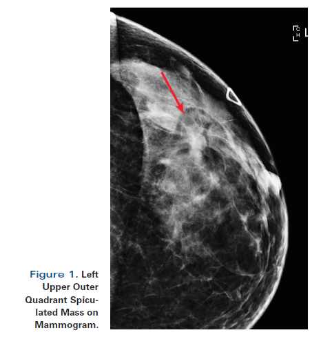


A 55 Year Old Woman With New Triple Negative Breast Mass Less Than 2 Cm On Both Mammogram And Ultrasound
In the left breast, the nerve generally exits the chest wall at the lateral border of the pectoralis minor, enters the posteriorlateral surface of breast at approximately the 4 o'clock position, and then traverses the glandular tissue to the inner areola along the 5 o'clock axis In the right breast, the nerve generally enters theLisa Jacobs, MD, Johns Hopkins breast cancer surgeon, and Eniola Oluyemi, MD, Johns Hopkins Community Breast Imaging radiologist, receive many questions about how to interpret common findings on a mammogram reportThe intent of the report is a communication between the doctor who interprets your mammogram and your primary care doctor However, this report is often I had a core biopsy on my breast this past Monday They called me with the results that I have a low grade Phyllodes tumor at the 12 o'clock position on my left breast I have been having other issues before the lump showed up two weeks ago I feel very tired, pain in my kidney position on both sides with no explanation, and now this lump with
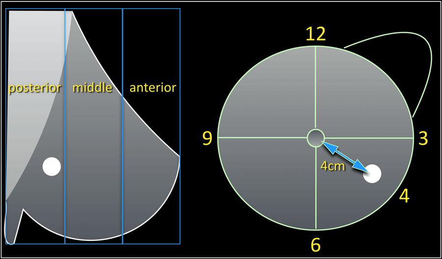


The Radiology Assistant Bi Rads For Mammography And Ultrasound 13


How To Self Examine Your Breast You Me Medicine
Breast cancer, ultrasonography The patient in Images 2628 also had a 7mmdiameter cyst at the 10o'clock position in the right breast My tumour was located at the 12o'clock position in my right breast Given the size and solid shape of the mass, my surgeon performed a lumpectomy The breast cancer surgery process often includes a sentinel lymph node biopsy (SLNB) (FCKING OUCH), where a chain of nodes are removed to test for cancer spread within the lymphatic system• there is a single tumor located at the 12, 3, 6, or 9 o'clock position on the breast Code the primary site to C509 when there are multiple tumors (two or more) in at least two quadrants of the breast Laterality Laterality must be coded for all subsites


Son 2147 Sonography Of The Breast Ppt Download
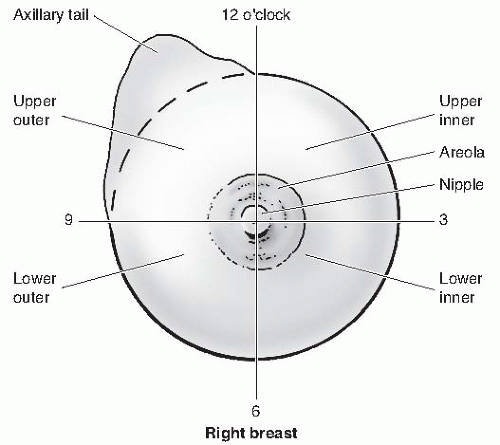


Breast Diseases Obgyn Key
O'Clock Positions and Codes Quadrants of Breasts 2 11 12 1 1 10 9 8 7 7 6 5 5 4 3 2 11 12 10 6 4 RIGHT BREAST LEF T BREAST UOQUIQ LOQ LIQ LIQ LOQ C502 C504 C502 C504 C505 C503 C503 C500 C501 SEER Program Coding and Staging Manual 07 C606 SiteSpecific Coding Modules Appendix C Abstract BACKGROUND Metaplastic carcinoma is a rare form of breast carcinoma that often is confused with other benign and malignant entities The diagnosis can be difficult to establish on both a cIn Breast Cancer ICD 9 diagnosis codes, there are no specific codes for the specific location of the breast cancer For example, ICD 9 Code 1742 for malignant neoplasm of upperinner quadrant of the female breast includes cancer on the right and/or left breast In Breast Cancer ICD 10 diagnosis codes, the positions of the cancer are specified


1
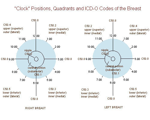


Epos Trade
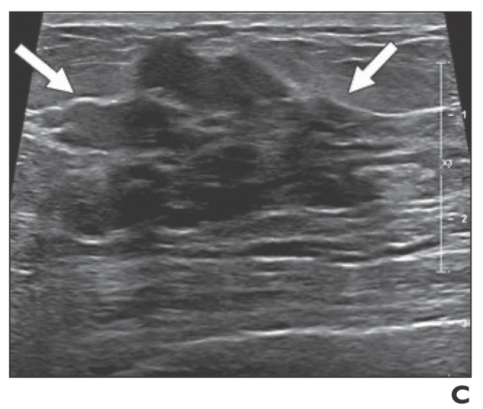
The right breast was normal to palpation Mammography revealed a solid, smooth nodule in the upper internal quadrant of the left breast (Fig 1) Ultrasound demonstrated an oval mass in the 10o'clock position of the left breast measuring 15×10 mm (Fig 2)Breast Cancer ICD10 Code Reference Sheet FEMALE Right C Malignant neoplasm of nipple and areola, right female breast C Malignant neoplasm of central portion, right female breast C Malignant neoplasm of upperinner quadrant, right female breast In the left breast there are two adjacent oval solid lesions measuring 5 and 65mm in diameter present in the 2 o'clock position In the 7 o'clock position there is a larger oval lesion measuring 10 x 5mm The appearance of each of the three lesions within the left breast is consistent, but not diagnostic of fibroadenoma


Cureus Invasive Lobular Breast Carcinoma Can Be A Challenging Diagnosis Without The Use Of Tumor Markers
/know-your-breast-tumor-size-4114640-FINAL-f17fb19bf9214d20937d07bd41524ac7.png)


Breast Tumor Size And Staging


Coding Breast Mass Becomes More Specific For Coders


Spie Org Samples Pm211 Pdf


Breast Mri Identified Lesions During Neoadjuvant Therapy Are Largely Benign


Contrast Enhanced Dedicated Breast Ct Detection Of Invasive Breast Cancer Preceding Mammographic Diagnosis Sciencedirect


Cancer Therapy And Oncology International Journal Ctoij


Cancer Therapy And Oncology International Journal Ctoij


National Program Of Cancer Registries Education And Training Series How To Collect High Quality Cancer Surveillance Data Ppt Download


Chapter 3 Lange Improved Flashcards Quizlet


Ultrasound Irregular Mass With Acoustic Shadow At Right 9 O Clock And Download Scientific Diagram


Osa Three Dimensional In Vivo Fluorescence Diffuse Optical Tomography Of Breast Cancer In Humans


Icd 10 Code For Breast Cancer At 12 00


Identification Of A Brca2 Mutation In A Turkish Family With Early Onset Breast Cancer Celik 18 Clinical Case Reports Wiley Online Library


Viral News Breast Anatomy Quadrants Breast Lymph Nodes And Lymphatic Drainage Clinical Role Kenhub Breasts Or Mammary Glands Are Capable Of Producing Milk In Females


Ultrasonography At The 12 O Clock Position Of The Left Breast Revealed Download Scientific Diagram


Factors Affecting Visualization Rates Of Internal Mammary Sentinel Nodes During Lymphoscintigraphy Journal Of Nuclear Medicine
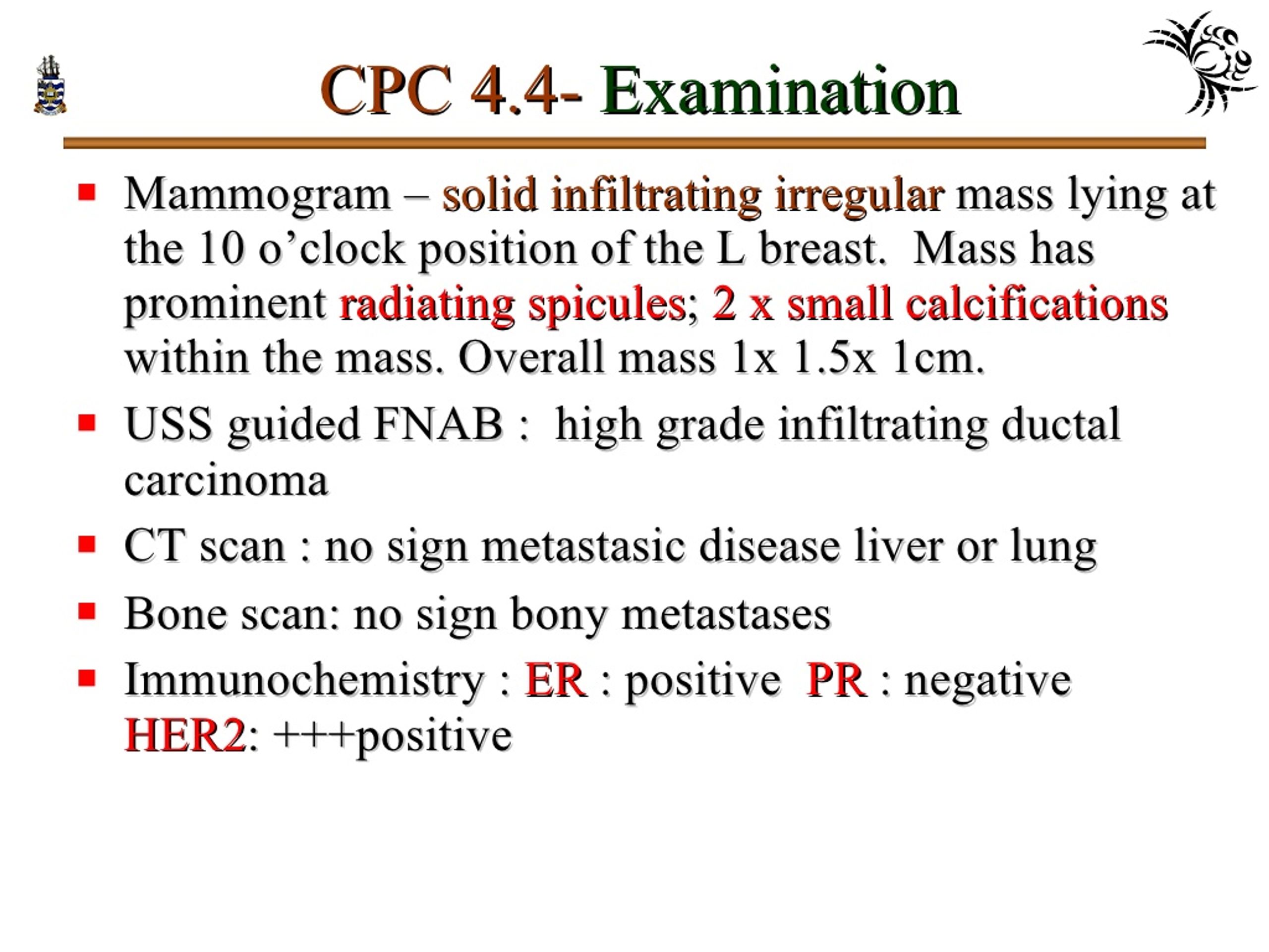


Ppt Pathology Of Breast Disorders Powerpoint Presentation Free Download Id
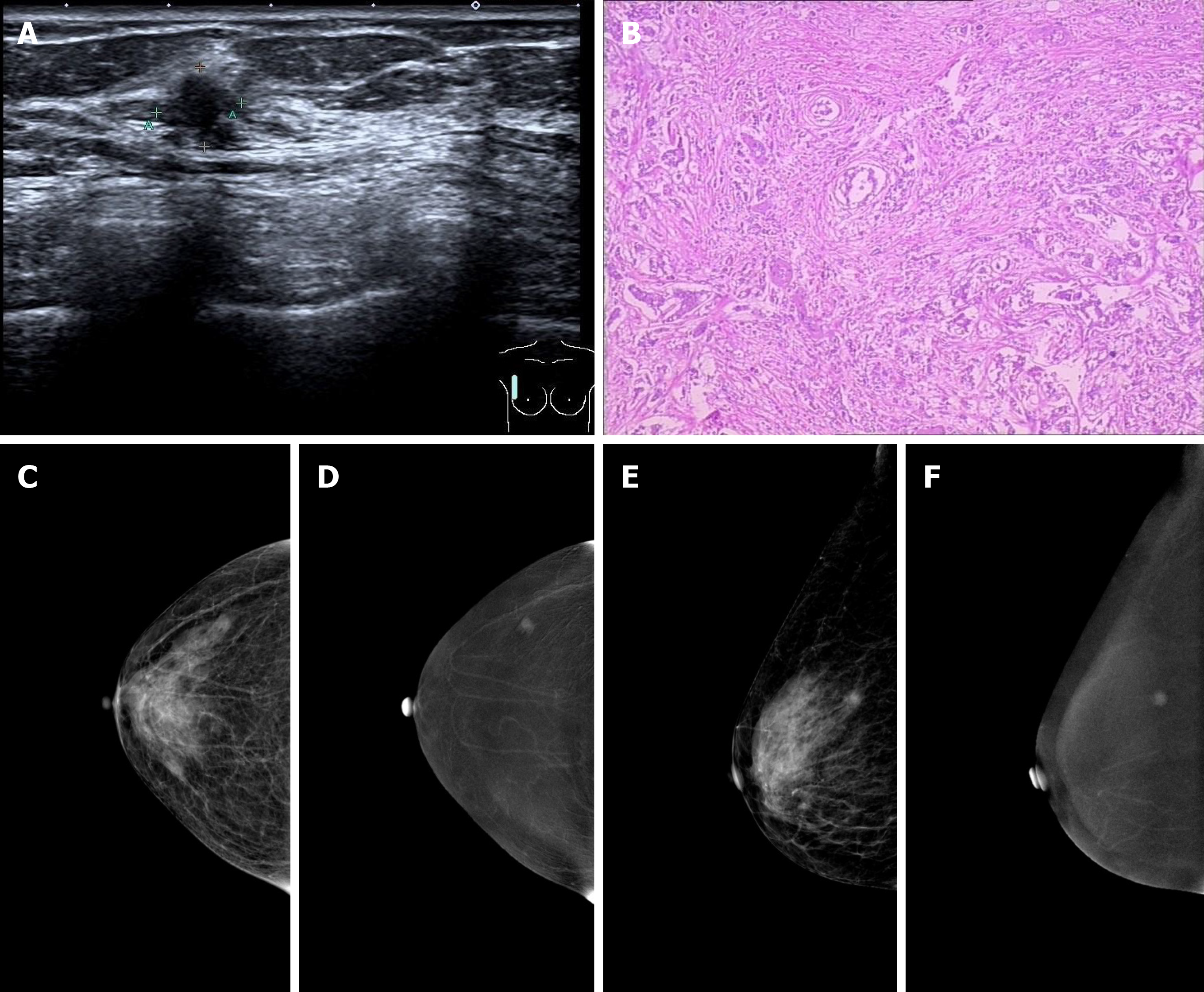


Primary Breast Cancer Patient With Poliomyelitis A Case Report


Breast 34 Fibrocystic Diseasechange Pathology Harmone Induced Acinar


Hypothetical Chief Complaint Ppt Download
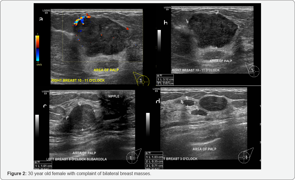


Cancer Therapy And Oncology International Journal Ctoij
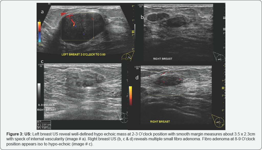


Cancer Therapy And Oncology International Journal Ctoij


Contrast Enhanced Dedicated Breast Ct Detection Of Invasive Breast Cancer Preceding Mammographic Diagnosis Sciencedirect
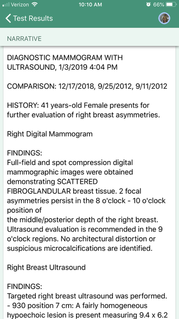


Breast Cancer Topic Interpreting Your Report


Breast Cancer Topic Quadrants Of Breast Cancer Survey Also


Nonvisualization Of Sentinel Node By Lymphoscintigraphy In Advanced Breast Cancer Topic Of Research Paper In Clinical Medicine Download Scholarly Article Pdf And Read For Free On Cyberleninka Open Science Hub
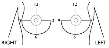


Breast Cancer Signs Symptoms And Understanding An Imaging Report Saint John S Cancer Institute


Gallery Formats Parvizbagloo


Journal Of Lancaster General Health Breast Cancer An Illustrated Case Study


Breast Cancer Topic Can Someone Help Me Understand My Results


Mammogram Revealing The Left Breast Mass A 5x4 4x1 9 Cm Irregular Download Scientific Diagram


Seer Training Quadrants Of The Breast


Quantitative Analysis Of Enhancement Intensity And Patterns On Contrast Enhanced Spectral Mammography Scientific Reports


Pik3ca Mutational Status Is Associated With High Glycolytic Activity In Er Her2 Early Invasive Breast Cancer A Molecular Imaging Study Using 18 F Fdg Pet Ct Springerlink


Imaging Findings For Malignancy Mimicking Nodular Fasciitis Of The Breast And A Review Of Previous Imaging Studies Topic Of Research Paper In Clinical Medicine Download Scholarly Article Pdf And Read For Free


Fcds Med Miami Edu Downloads Naaccr Webinars Breast Breast final Pdf


Seer Cancer Gov Manuals 21 Appendixc Coding Guidelines Breast 21 Pdf


Recent Comprehensive Review On The Role Of Ultrasound In Breast Cancer Management Leaders In Pharmaceutical Business Intelligence Lpbi Group


1


Secretory Breast Carcinoma A Report Of Three Cases And A Review Of The Literature


Www Bjcancer Org Sites Oldfiles Library Userfiles Pdf 137 Pdf


Osa Three Dimensional In Vivo Fluorescence Diffuse Optical Tomography Of Breast Cancer In Humans


Print Anatomy Physiology And Pathology Of The Breast Mammography Flashcards Easy Notecards


Diagnostic Breast Imaging Clinical Breast Imaging A Patient Focused Teaching File 1st Edition


Distribution Of Breast Cancer Locations In The Breast According To Download Scientific Diagram
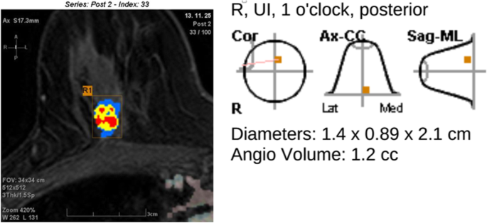


Preoperative Tumor Size Measurement In Breast Cancer Patients Which Threshold Is Appropriate On Computer Aided Detection For Breast Mri Cancer Imaging Full Text


Diagnosis And Management Of Benign Breast Disease Glowm


Bilateral Breast Cancer Different In More Ways Than One


Www Birpublications Org Doi Pdf 10 1259 Bjr


Coding Breast Mass Becomes More Specific For Coders


Http Static pc Com A3c7c3fe 6fa1 4d67 8534 A3c9c15fa0 C7b39f96 0935 4e94 84b8 C4a6d9d86abd 3739cb 8b1c 4e15 6b E6192eec5078 Pdf


O Clock Position Of A Lesion On A Right Breast As Identified By Download Scientific Diagram


Normal Ultrasound Protocol Breast Ppt Video Online Download
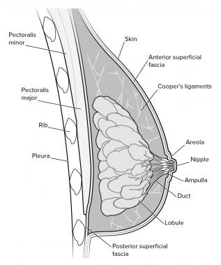


Mammography In Breast Cancer Background X Ray Mammography Ultrasound


Individualized Treatment Analysis Of Breast Cancer With Chronic Renal Ott


Healthy Breast With Ultrasound Radiology Key


Diagnostic Breast Imaging Clinical Breast Imaging A Patient Focused Teaching File 1st Edition


Www Thieme Connect De Products Ebooks Pdf 10 1055 B 0034 Pdf
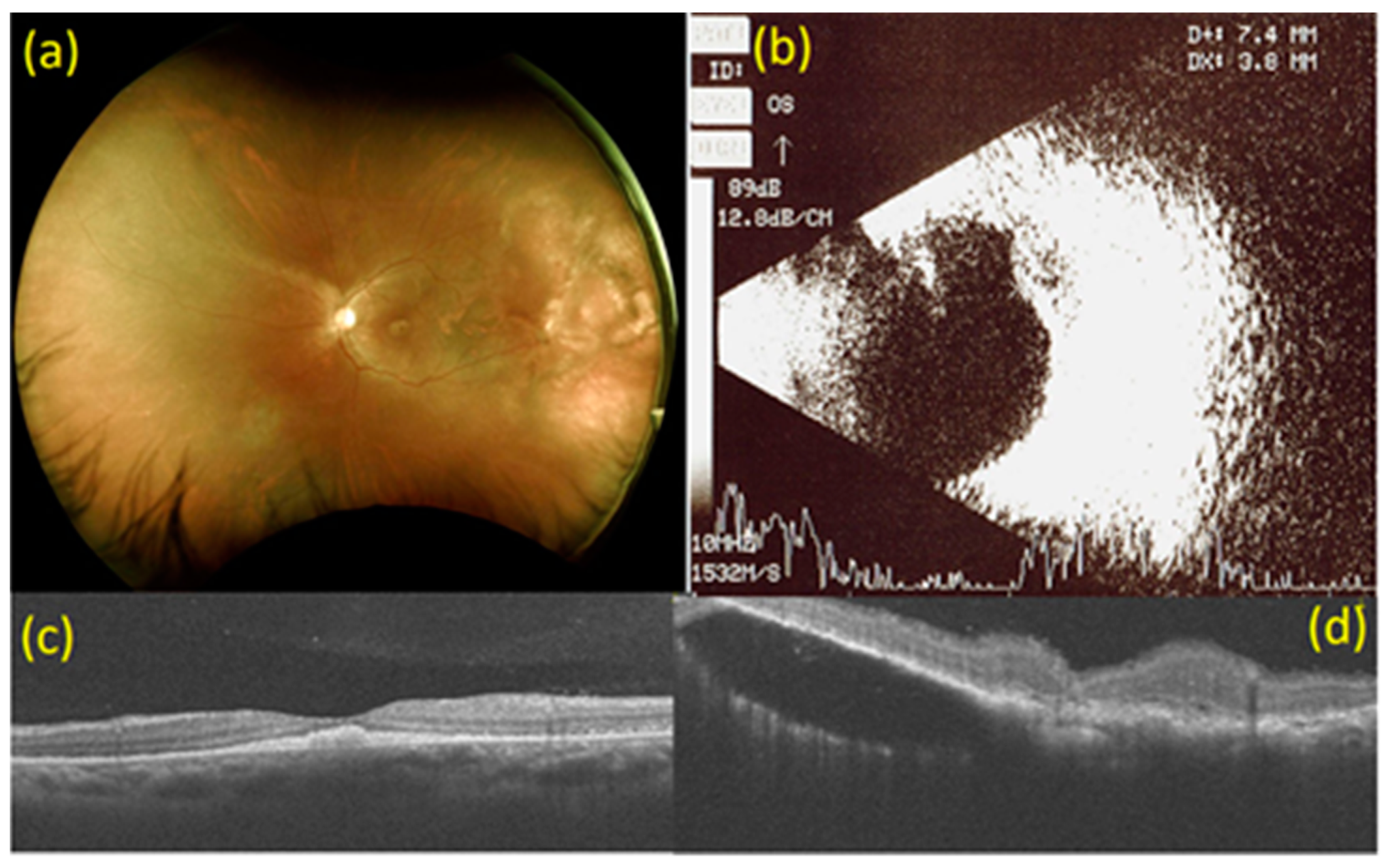


Medicina Free Full Text Adjuvant Intravitreal Bevacizumab Injection For Choroidal And Orbital Metastases Of Refractory Invasive Ductal Carcinoma Of The Breast Html


Lange Q A Chapter 3 Anatomy Physiology And Pathology Of The Breast Flashcards Quizlet


Coding Breast Mass Becomes More Specific For Coders
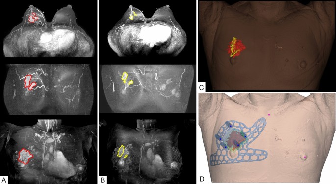


Usefulness Of 3d Surgical Guides In Breast Conserving Surgery After Neoadjuvant Treatment Scientific Reports


Diamond Mammoplasty As A Part Of Conservative Management Of Breast Cancer Description Of A New Technique Sciencedirect
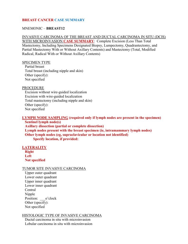


Breast Invasive Carcinoma
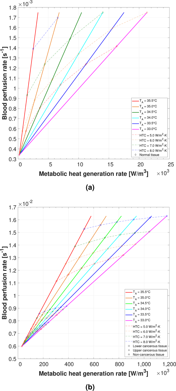


Determining The Thermal Characteristics Of Breast Cancer Based On High Resolution Infrared Imaging 3d Breast Scans And Magnetic Resonance Imaging Scientific Reports


Experts To Consider All Aspects Of Breast Ultrasound European Society Of Radiology
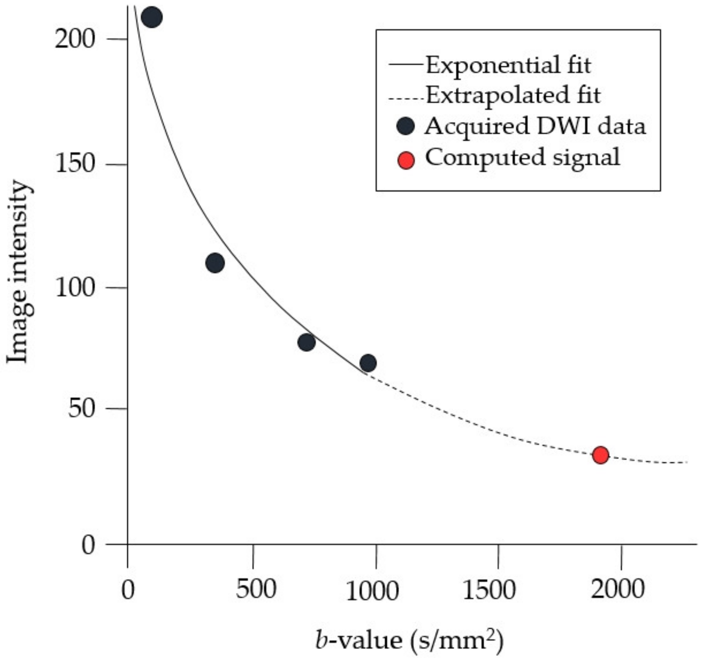


Diagnostics Free Full Text Clinical Feasibility Of Reduced Field Of View Diffusion Weighted Magnetic Resonance Imaging With Computed Diffusion Weighted Imaging Technique In Breast Cancer Patients Html


26 Year Old With Palpable Mass Near The 12 00 O Clock Position Of The Download Scientific Diagram


1


Ultrasonography At The 12 O Clock Position Of The Left Breast Revealed Download Scientific Diagram


Pdf Electrical Impedance Spectroscopy For Breast Cancer Diagnosis Clinical Study Semantic Scholar


Distribution Of Breast Cancer Locations In The Breast According To Download Scientific Diagram


Nucleus Iaea Org Hhw Radiology Clinical Applications Specialities And Organ Systems Breast Imaging Guidelines And Literature Suggested Articles Reading A Mammogram Pdf


Lactational Breast Changes Lobular Hyperplasia Mimicking Masses How Can We Differentiate From True Pathological Masses


Epos
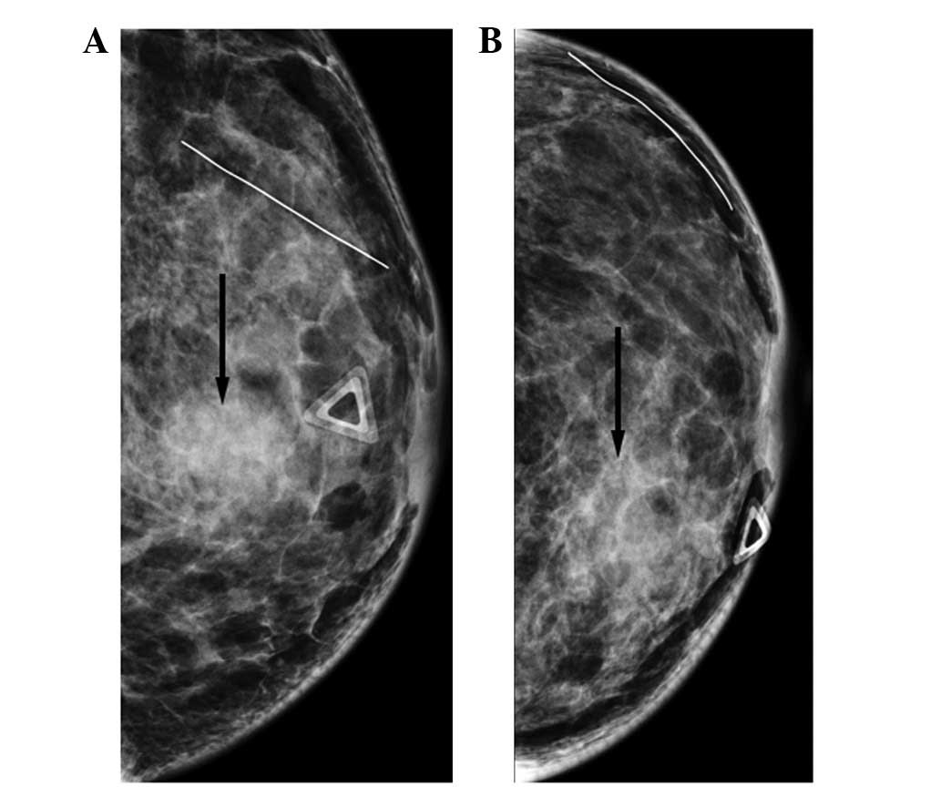


Breast Metastasis From Nasopharyngeal Carcinoma A Case Report And Review Of The Literature


Www Biobran Org Uploads Case Study 3a1d32a1d152e7f1b1c5818d322b81ac Pdf
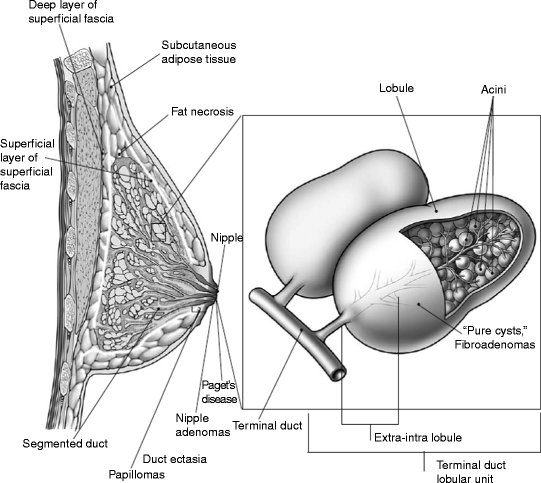


Breast Cancer Springerlink


Ispub Com Ijanp 12 1 2947


0 件のコメント:
コメントを投稿